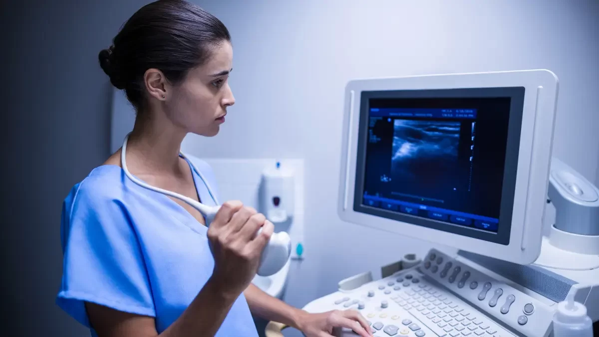
Ultrasound / Digital X-Ray are essential imaging techniques used in medicine to provide detailed images of the internal body. These non-invasive procedures are widely used for diagnosing and monitoring various conditions. Ultrasound uses sound waves to create images of organs and tissues, while Digital X-ray uses radiation to capture detailed images of bones and tissues. Both techniques are efficient and have revolutionized the ability to detect and treat health conditions earlier. They are applied across various fields like orthopedics, cardiology, and gynecology, allowing healthcare professionals to make timely, accurate treatment decisions and improve patient care. These imaging methods help in quick diagnosis, which is crucial for implementing appropriate treatment and avoiding complications.
They also play a key role in assessing disease progression, ensuring optimal care. With advancements in technology, Ultrasound, Digital X-ray, Echo, TMT, and PFToffer enhanced accuracy and capabilities in healthcare.
What is Digital X-ray?
A Digital X-ray is an advanced imaging method that uses low levels of radiation to capture detailed internal images. Unlike traditional X-rays that use film, Digital X-rays utilize digital sensors to convert X-ray data into digital images. These images are then displayed on a computer screen for further examination. Digital X-rays offer faster image processing, clearer resolution, and allow for image enhancement. They are commonly used for diagnosing bone fractures, tumors, infections, and other medical conditions. This technology is particularly valuable in emergency care, orthopedics, and dental applications. It helps healthcare professionals identify issues quickly and accurately, which is essential for timely interventions and better treatment outcomes.
- Quick Image Processing: Results are available instantly, enabling healthcare providers to make timely decisions.
- Improved Image Quality: Digital X-rays offer higher resolution and allow image enhancement for better diagnosis.
- Lower Radiation Exposure: Compared to traditional X-rays, digital images require significantly less radiation.
X-Ray Scan Procedure
The Digital X-ray procedure is straightforward and non-invasive. Patients are typically asked to change into a gown and remove any metal items such as jewelry. The patient is then positioned in front of the X-ray machine, and depending on the area being examined, the patient may either stand, sit, or lie down. The technician will direct the X-ray machine to the correct angle and may ask the patient to hold still or briefly hold their breath while the image is taken. The process is quick, usually taking only a few minutes. Occasionally, multiple views may be needed for a comprehensive image. This simplicity and speed ensure that the patient is not kept waiting for long, and the procedure remains hassle-free.
- Simple and Quick: The procedure takes only a few minutes, with minimal discomfort for the patient.
- Precise Positioning: The technician ensures proper alignment to capture the most accurate images.
- Non-invasive: There’s minimal patient interaction with no invasive procedures required.
Uses of X-Ray Scan
Digital X-rays are used to diagnose a variety of conditions affecting bones, joints, and internal organs. In orthopedics, they are commonly used to detect fractures, joint dislocations, and degenerative conditions like arthritis. In dentistry, X-rays are used to identify cavities, infections, and gum disease. Digital X-rays are also valuable for diagnosing lung issues such as pneumonia or tuberculosis and assessing heart-related conditions. Additionally, they play a crucial role in emergency care, providing immediate insights into trauma injuries, fractures, and other acute health concerns. Their ability to provide fast, clear images makes them essential in a variety of medical settings, helping physicians make informed decisions quickly and accurately.
- Bone and Joint Assessments: Digital X-rays are extensively used in orthopedics to evaluate bone fractures, joint dislocations, and degenerative diseases.
- Dental Diagnostics: Dental X-rays are crucial for detecting cavities, gum disease, infections, and planning orthodontic treatments.
- Internal Organ Imaging: Digital X-rays are valuable for diagnosing conditions affecting the chest, abdomen, and other internal organs, providing insight into issues like infections, tumors, and more.
Benefits of Digital X-ray
Digital X-rays offer numerous advantages over traditional film-based X-rays. The primary benefit is the speed; digital images are processed and displayed almost instantly, allowing doctors to review them quickly and make prompt decisions. The image quality is superior, as digital X-rays provide high-resolution images that can be easily enhanced for better clarity. Additionally, Digital X-rays are more eco-friendly, as they eliminate the need for chemical processing and film storage. They also use less radiation than conventional X-rays, making them safer for patients, particularly those requiring frequent imaging. The reduction in radiation exposure is one of the most significant benefits, especially for children, the elderly, and patients who need repeated scans.
- Faster Results: Digital X-rays provide immediate images, enabling faster diagnosis and treatment decisions.
- Superior Image Quality: Enhanced images provide clearer details, which aids in accurate diagnosis.
- Reduced Radiation: Digital X-rays use significantly less radiation than traditional X-ray technology, making them safer for patients.
Preparation
Preparing for a Digital X-ray is simple. Patients may be asked to change into a hospital gown to avoid interference from clothing. It is important for patients to inform the medical staff if they are pregnant or might be pregnant, as radiation can pose risks. Depending on the area to be examined, the patient may need to avoid eating or drinking for a few hours before the procedure. In the case of an abdominal or chest X-ray, certain medications or supplements may need to be temporarily stopped, but this will be discussed by the healthcare provider beforehand. Preparation is generally minimal and straightforward, making it a quick and hassle-free process for most patients.
- Wear Appropriate Clothing: Patients should wear a gown, and remove jewelry or metal accessories.
- Avoid Eating or Drinking: For abdominal X-rays, patients may need to fast before the procedure.
- Inform the Technician: Let the medical staff know if you’re pregnant or could be, to ensure safety during the procedure.
Conclusion
Digital X-ray is a crucial diagnostic tool that offers fast, clear, and accurate imaging for a wide range of medical conditions. Its efficiency, high-quality images, and reduced radiation exposure make it a preferred choice for many diagnostic needs. From diagnosing bone fractures to identifying internal organ issues, digital X-rays provide a comprehensive solution for examining the body’s internal structures. At Prime Indian Hospital, we utilize the latest Digital X-ray technology to ensure that our patients receive accurate and timely diagnoses. Whether for routine examinations or in emergency situations, Digital X-ray remains a reliable, non-invasive option that aids in improving patient care and outcomes.


